By Eva Briggs, MD
The risk of developing condition increases with obesity and with increasing age.
Recently I saw a patient with a previously undiagnosed condition called Charcot foot. This is a foot deformity occurring in patients with diabetes or others with decreased sensation in their feet.
The path to Charcot foot begins when diabetes damages small blood vessels.
This impedes circulation to the nerves, causing nerve damage called neuropathy. Patients with neuropathy lose the ability to detect cuts or other damage to the foot. Poor circulation also impairs the body’s ability to heal cuts, injuries, and infection. The foot is not only at higher risk for injury and infection, but heals these problems slowly. If you’re experiencing these symptoms, it’s crucial to seek “neuropathy treatment near me” to manage the condition effectively.
This is an important reason for patients with diabetes to work with their doctors to control their blood sugar as best as possible. They should inspect their feet every single day to discover cuts, blisters or other problems right away. Clean feet, clean socks and well-fitting protective footwear are essential.
Charcot foot — also called Charcot arthropathy — develops only when neuropathy is present. The foot becomes injured, and the patient does not feel or sense the injury. The injury can be from obvious trauma, such as a fall, a twist, dropping something on the foot, or hitting the foot against an object. Often the injury is a series of small, undetected, cumulative microtraumas.
In a normal foot, abnormal forces or weight distribution is detected right away, and leads to automatic correction by adjusting the stance.
People without neuropathy do this all day, every day, automatically without conscious awareness. But diabetic patients lack this ability to auto-adjust. These small traumas accumulate and lead to fractures and other injuries.
Without the ability to sense pain, the diabetic patient continues to walk on their injured foot. The injuries worsen leading gradually to foot deformity.
Less than half of patients with a Charcot foot even recall any injury. The risk of developing Charcot foot increases with obesity and with increasing age.
The deformity in Charcot foot makes fitting into normal shoes difficult or even impossible. The foot and ankle may become unstable, preventing normal walking. Most dangerous of all, the deformed bones in Charcot foot may push against the skin leading to a high risk of ulcers. Skin ulcers are at high risk of infection. Poor circulation makes healing of the ulcer and any associated infection difficult. Patients with infected Charcot foot ulcers have an incredibly high 50% risk of an amputation.
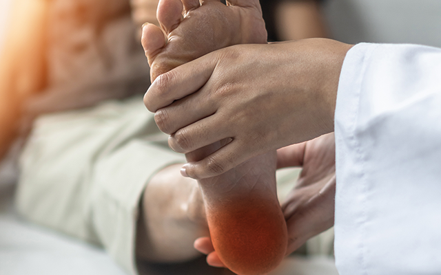
The first sign of a developing Charcot foot is swelling, usually painless. This often manifests as trouble fitting into one’s shoes. Another sign is redness and warmth, the body’s inflammatory response to injuries in the foot. Although this resembles infection, the risk of infection is low when the skin is intact. The redness and warmth of Charcot foot usually improve with elevation. Redness and warmth are less likely to resolve with elevation in infection.
To diagnose Charcot foot, your doctor will review your history and examine your foot. Tests that may help with the diagnosis include X-ray, CT, MRI or bone scan. These tests help distinguish Charcot foot from infection and aid in planning the best treatment.
The goal of treatment is to create a foot that can rest flat on the floor, is without ulcers, and if possible, can use a commercially available diabetic shoe.
Nonsurgical treatments for early-stage Charcot foot include casts or boots to allow the bones to heal in a stable position and to prevent further deformity. Casts can reduce swelling and protect the bones. While wearing a cast, the patient will likely need to avoid weight bearing and use crutches, a knee scooter or wheelchair.
Patients unable to wear regular over-the-counter shoes on a deformed foot may need custom shoes, braces or orthotics.
When these treatments are insufficient, surgery may be an option. Some types of surgery include debridement (cleaning) foot ulcers, lengthening the calf muscle or Achilles tendon to relieve pressure points on the foot and removing bony prominences that push on the skin creating the risk of ulcers. Sometimes more complex surgeries are required to correct the deformity. Due to the complexity of the surgery combined with diabetes, these surgeries carry a higher risk of complications, infection and amputation than routine foot and ankle surgery. For this reason, it is imperative to strive for the best possible blood sugar control and to avoid weight bearing until OK’d by the surgeon.
If you have diabetes, your best bet to protect your feet is controlling your diabetes, checking your feet daily and wearing strong supportive footwear. If you do notice a foot problem, don’t delay in seeking medical care.

Eva Briggs is a retired medical doctor who practiced in Central New York for several decades. She lives in Marcellus.